Courtesy of Intermountain Medical Imaging, Boise, Idaho. All rights reserved.
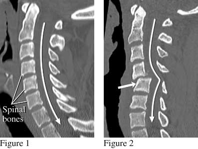
The side view of the spine (figure 1) shows the normal position of the spinal bones. The spinal cord (not visible in this image) follows the path of the long white arrow. Figure 2 shows shifted spinal bones (short thick arrow) from a fracture that damaged the spinal cord. This person was paralyzed after being ejected during an automobile crash while not wearing a seat belt.
Current as of: July 26, 2023
Author: Healthwise Staff
Clinical Review Board
All Healthwise education is reviewed by a team that includes physicians, nurses, advanced practitioners, registered dieticians, and other healthcare professionals.